Episode 68
Picture This: Diagnostic Imaging for the General Practitioner
With Marc Seitz
MIDWEST VETERINARY CONFERENCE PREVIEW SERIES See All Episodes »
Radiology might be intimidating for the average small animal practitioner, but it doesn’t have to be! There’s no need to get board certified and become a specialist to use imaging as a diagnostic tool in your practice. All you need is a little continuing education, some pointers, a few tips and tricks, and you’ll be on your way to using radiology like a pro.
Dr. Marc Seitz (who is, in fact, a board-certified veterinary radiologist) has a goal: To translate complicated medical practices and scientific literature into useful clinical skills that all practitioners can use in their everyday practice. In this episode, is giving listeners a sneak peek into his Pet Animal Radiology sessions at the 2023 Midwest Veterinary Conference.
Episode Guest
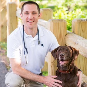
Marc Seitz
DVM, DACVR, DABVP (CANINE/FELINE)
An associate clinical professor of diagnostic imaging at Mississippi State, Dr. Seitz is a board-certified radiologist and ABVP diplomat. His teaching style earned him the Zoetis Distinguished Veterinary Teaching Award in 2016. | Learn More »
Registration for the 2023 Midwest Veterinary Conference is open! Featuring 300+ hours of live and on-demand CE in 25 tracks, 75 expert speakers, and more than 100 exhibitors, this is another event you won’t want to miss!
Photo by Tima Miroshnichenko on Pexels
Transcript
Mia Cunningham: Thanks for joining us, Dr. Seitz.
Marc Seitz: I love it. I am so excited! This is actually going to be my first time attending the conference, and I’m very stoked to get back to my birth state. I actually have an Ohio Social Security number, so this is going to be good. I was born in Akron, Ohio, and I have not been there for a while.
Krysten Bennett: Awesome! O-H!
MS: Yea!
MC: I-O!
MS: I’m rusty.
KB: Well, you’ll fall back into it. It’s like riding a bike. Since you’re in Ohio native, we don’t have to warn you about the weather, do we? Because Columbus in February…
MC: Can be tricky.
KB: It is tricky. It can kind of go either way.
MS: I’m prepared to have to drive if flights get canceled. You guys don’t get the lake effect snow down in Columbus, but in Akron, we were always dealing with the lake effect snow. It was awful. So awful.
KB: We’re lucky that we’re far enough away. I mean, I think we’re lucky. I’m not a big snow person. But anyway, we’re here to talk about the MVC!
MS: I think it’s neat that you guys do this. I’ve never done this for a conference before, so for being a regional conference, I feel like you’re kind of ahead of the game on most other regional conferences. I’m serious! This is really good. I’ve only ever seen the hype stuff done by national conferences like Western and North American—VMX now, but you know what I mean. This is the most well-run regional conference I’ve seen yet. So, kudos.
MC: Thank you so much.
KB: We appreciate it, and we’ll continue showing off to you in the coming months. But in the meantime, we want to hear about what you’re bringing to MVC. So can you start us off by telling our listeners about you and share a little bit about your background?
MS: I am Dr. Marc Seitz. I am currently an associate clinical faculty member at Mississippi State University and I’m a radiologist. Prior to my life in academia, I was actually in private practice. When I graduated, I spent three years as a full-time general practitioner and a part-time emergency vet, and then for five years after that, did full-time ERand part time GP, spent a few years as an emergency faculty, and then eventually embraced my passion for imaging and pursued a second specialty in diagnostic imaging.
MC: Awesome. So we’re excited to have you join us as a speaker for the 2023 Midwest Veterinary Conference. If you could give our listeners just a peek into the sessions that you’ll be providing and a little bit about what people can expect to learn.
MS: So I was selected to pick six topics—obviously all imaging heavy, but what’s fun is I like to make imaging clinically relevant. My goal is to make imaging into something that can be used the next day. Because we all go to conferences, not to be nerds, but to help our patients. So, all the talks were written from the perspective of things that I wanted when I was a general practitioner, either in primary care or emergency medicine.
So for the first talk, we’re going to get into one of the most common things we see, and that’s diagnosing gastrointestinal obstructions. Even now I, like the listeners, to hear as a radiologist, my stomach still turns and knots because of the importance of that call. And so what we’re going to do is just throw a whole bunch of cases up and go through them and talk about all the little nuances we look for, not as a radiologist but as a general practitioner. What can we do to maximize the yield of radiographs? Because I think they are still valuable and ultrasound should not be replacing abdominal radiographs. We can still make a diagnosis of obstruction.
We’re then going to segue and talk about some scarier abdominal diseases. We’re going to talk about torsions, the most common being gastric dilatation and volvulus, but we’re going to spend some time talking about the less common ones. And also, again, tips and tricks we can do to increase the likelihood that we make this crucial call correctly. Because these are all emergencies that, with prompt surgical intervention, do pretty well, but without it, we can unfortunately impact the prognosis negatively.
We’ll then transition to one of my favorite diseases—not for the patient, but just it’s something that I have a very strong interest in, and that’s blocked cats. We see it all the time, right? And so I want to make certain that we can cover all the wonderful imaging things that, again, we can do in a general practice setting to increase the likelihood that we’re going to find out what’s causing them to be obstructed. We’re going to talk about a few new articles that have come out. For example, it was a surprise to me when I read it, but blocked cats, male cats, actually can have an os penis, and that os penis can actually mimic stones or crystal plugs. So we’re going to talk about some fun updates like that that I think will be very useful. We’ll also talk about contrast studies because they’re super easy and shouldn’t be just done by radiologists.
Then we’re going to move into my favorite imaging modality, which is ultrasound. Absolutely love it. More and more general practitioners are doing it. They should be, whether we’re doing it in a point-of-care fashion or doing it more diagnostically, we want to make certain that we’re utilizing that ultrasound. So we are going to first talk about getting started with ultrasound. I’m going to talk about basic knobs. You play with what the image looks like, what makes the image, a couple of different machine types based on your goals and what you’re trying to achieve.
We’ll then talk about the most common utilized ultrasound technique, which is point-of-care ultrasound. Some of our users or attendees may know this as FAST, but FAST just really doesn’t encompass all that we use it for. It’s really a bedside point-of-care test now, and we’re going to talk about going beyond a basic FAST scan or a basic point-of-care ultrasound, and some of the extra things we can look for in the abdomen that can really make a difference in how we work that patient up. For example, one thing that has become a hot topic is the gallbladder halo. And there’s been a lot of attention recently on anaphylaxis and gallbladder halo or gallbladder thickening. But we’re up to 30 or 40 different causes. And based on some new research that I’m going to share, believe it or not, we can induce a gallbladder or halo just by choosing a certain sedative drug. So I want to make certain that we’re not misdiagnosing a normal change that we see with sedation for something more scary like anaphylaxis, gallbladder disease, pericardial fusion, something. So that’s going to be an exciting talk.
And then finally, we need to give our cats some more attention. And I’m going to give a nice overview of the basic feline abdomen and how their abdomen differs from dogs anatomically. We’re going to talk about how they get these really cute bilobed gallbladders. We’re going to talk about how their liver looks different. We’re going to talk about their bile duct, their pancreas. For example, we see the left limb of the pancreas more commonly in the cat than the dog, whereas in the dog we see the right limb. So we’re going to go over all the species differences that, if we’re applying that same dog algorithm, will be wrong sometimes. So I want to make certain that we know all the normal feline species differences for those general practitioners that are doing complete diagnostic scans.
KB: So it sounds like your sessions aren’t necessarily geared towards somebody who is a specialist, that it’s something that any general practitioner can use.
MS: Absolutely. That is actually the goal for 90% of my CE. It is geared toward general practitioners of all skill sets. I really hope new graduates and people new to imaging or rusty on imaging come out. But also I bury at least three to five fun updates for all my really skilled general practitioners who might look at it and go, oh, FAST, I already know how to do that. Or torsions: yeah, I know everything there is about that. My goal is that even for those, they leave with three to five points. So if we do have some specialists, I do think they’ll glean a few things.
But I am not here to lecture to radiologists or the surgeons or internists. I want this information to be valuable to the general practitioner. And more importantly, I want them to go next week, enter their clinics, and I want to get flooded with emails that said, “Dr. Seitz, I learned X, used it on a patient, and it helped them.” If I’ve done that, I’ve done my job.
MC: Awesome. Now, if any of our attendees would like to connect with you or have access to you online, where can they find you?
MS: Yeah. I think the two easiest ways are going to be my email address, which is Marc.Seitz@msstate.edu, and I respond to my email usually within about 72 hours. And then the other is I’m on LinkedIn. I love LinkedIn! It is probably the most up to date bio I have. Sometimes university websites are a little slow to update, but you can also find me on the Mississippi State University website, but they still have me a little bit behind with where I’m at in my career. So find me on LinkedIn and you can email me as well.
MC: Was there anything else you’d like to share with us before we let you go?
MS: Conferences are one of my favorite things in the world. I am looking forward to meeting all of our veterinarians in Ohio and the greater states that surround. It’s going to be fun to be back in my birth state. And one of the things I like is meeting people, hearing their stories. So come up, chat with me. And of course, if I have any former students or trainees, please come up and say hi.
KB: That’s great. MVC is more than just amazing continuing education, folks. We’ve got networking, we’ve got exhibits, we’ve got podcasts!
MS: Where do I find these podcasts? Where are they going to show up?
KB: They’ll be on FullyVettedPodcast.com, Apple, Spotify, all the usual podcast places. Subscribe! Never miss an episode!
MS: I will. Well, you all thank you for your time.
KB: Thank you so much.
MC: Bye bye.