Episode 69
Veterinary Detectives: Using Cytology to Solve Medical Mysteries
With Kate Baker
MIDWEST VETERINARY CONFERENCE PREVIEW SERIES See All Episodes »
Like many of this higher sciences, cytology can be intimidating to general practitioners. How do you get a good sample? What are you looking for? How do you use the results? For some, it’s just easier to refer clients to specialists. But it doesn’t always have to be difficult!
On this episode, board-certified clinical pathologist Dr. Kate Baker explains her passion for cytology—and reassures listeners that there are tips and tricks that can help you solve the mystery. She’s offering a sneak peek into her sessions at the 2023 Midwest Veterinary Conference, which will explore practical techniques for getting good samples, how to identify common diseases cytologically, and pitfalls to avoid.
Episode Guest
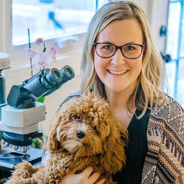
Kate Baker
DVM, M.S., DACVP
Dr. Baker became board certified in Veterinary Clinical Pathology in 2016 and currently works as an educator, diagnostician, and consultant. She is founder and CEO of VetHive. | Learn More »
Registration for the 2023 Midwest Veterinary Conference is open! Featuring 300+ hours of live and on-demand CE in 25 tracks, 75 expert speakers, and more than 100 exhibitors, this is another event you won’t want to miss!
Photo by Artem Podrez on Pexels
Transcript
Krysten Bennett: Hey, Dr. Baker. Thank you for hopping on here with us.
Kate Baker: Of course! Hopefully there won’t be too much barking or background noise, but I know that that’s kind of just par for the course on these things.
Krysten: Yeah. Right. We kind of expect it. How many dogs do you have?
Dr. Baker: Two. And they’re little yappy things, a cavapoo and a Chihuahua. So, like, you know, at any given moment, all hell could break loose, so it’s always a fun game.
Krysten: Yeah, I’m familiar. I brought my pibble to work today, and she’s been lying in the hallway, staring at the door, intermittently barking her head off at potential threats like passersby and mail people.
Dr. Baker: I have a livestock guardian dog too. It’s a Great Pyrenees. It’s his name Sheriff Gus, and he does the same, but he’s outside.
Krysten: Sheriff Gus. I love that.
Dr. Baker: Yeah. He protects our chickens and make sure they don’t get picked off by foxes.
Krysten: He’s got a very important job.
Dr. Baker: Yes, he does.
Krysten: And I think our audience — (dog barking in background) — understands.
Dr. Baker: Right. Exactly. One second, let me lock the dogs in the bedroom. They’re doing the walk of shame. They’re like, what did we do?
Krysten: I think the walk of shame means something different for them than it does for humans.
Dr. Baker: (laughs) Yeah, well, sure. All right. Okay. We are good. So whenever you’re ready.
Krysten: Okay. Let’s just start out with an introduction. Can you tell us a little bit about yourself and how you got into veterinary medicine?
Dr. Baker: Sure, sure. My name is Dr. Kate Baker. I’m a board-certified veterinary clinical pathologist. How I got into veterinary medicine. Well, how far back do we want to go here? I would not say that I was one of those kids that wanted to be a vet, but I started having an interest in, and I was really drawn to, veterinary medicine in high school, so, I mean, I was still a kid. I got a job at a local veterinary clinic as a kennel assistant, and I was very much a science geek. Even at that point in my life, I loved the melding of science and animals. I didn’t really know a ton about it—obviously, I was only 16—but I felt drawn to it.
So I went into college pre-vet. Went all through undergrad pre-vet. I dappled in thinking about other things, maybe med tech or human medicine, but kept getting drawn back to veterinary medicine. It was towards the end of my undergrad years that I actually started to develop a really strong interest in the higher sciences: Those upper-level biology classes, micro and embryology, histology, molecular biology. I was, like, really into those. I mean, they were tough, but I was really into them. I kind of had this moment where I thought, “Well, how in the world do I combine this love of these really intense sciences with veterinary medicine?” Because, I mean, those are obviously part of veterinary medicine. But I loved the intricacy of the microscopic world, and so I looked into it and realized that veterinary pathology was a specialty within veterinary medicine.
So even before going to vet school, I knew I wanted to be a pathologist, and that was my goal the whole time through vet school, was to be a pathologist. I never really had any intention to practice. I was pathology all the way through. But I did do an internship after vet school, and as most internships are, it was very intense, and it was good for me. I was glad to have that experience in the clinician’s seat, treating patients and being part of that process and understanding how those conversations go with owners and all those things. But it pretty much solidified for me that it was not the route I wanted to take long term. And then I went on and did my residency and pathology.
Krysten: And where are you working now?
Dr. Baker: I have a pretty interesting and non-traditional career now. So after my residency, I went on and did almost five years at a large diagnostic lab doing high-volume diagnostics, and I really enjoyed it. I got a ton of experience with that, seeing so many cases, but I really missed teaching. I really missed the creative part of me. I tend to have a bit of a creative side that I want to make things and teach, and I just felt like there was more I wanted to do.
So now what I do is, I work for myself. I have my hands in several different things. I do a lot of different things, which makes my days very interesting. I provide continued education courses for veterinarians and veterinary technicians online, and so I’ve been doing that for about four years now. I also have a consultation service. It’s an app-based tele-cytology service called Pocket Pathologist where I read cases. Again, I created this, and I am the sole pathologist of it at this time, but that will change.
And then I have something exciting coming up, too, that I’m working on in January that I can’t tell about yet. But just to give a little teaser, by the time we’re at Midwest, it’ll be out there. It’s going to be a very exciting community for veterinarians. So that’s just a little teaser. But I also do a lot of speaking, a lot of continued education type things, and obviously conference speaking.
Krysten: And that’s the perfect segue into why you’re here today! Dr. Baker is going to be speaking at the 2023 Midwest Veterinary Conference, and she’s going to be speaking about cytology. Can you give us a brief overview of what your subjects are?
Dr. Baker: Yes. So as a clinical pathologist, we have a couple of different areas of expertise, but my particular strong area of interest is cytology. So I do a ton of teaching on cytology, and that’s what my sessions will be focused on. I have multiple sessions. I think I have six, and it’ll all be focused on psychology. For the general practitioner, basically. How can you utilize cytology in practice? Where do you even begin with cytology? Cytology is one of those subjects that, I think, historically were not taught a lot about in school. There’s just only so many things that the vet schools can fit into the curriculum, and cytology just tends to not be heavily focused, which is neither here nor there. It’s just a fact for most schools.
And so I think that there’s a lot of discomfort for, really, all veterinarians. I see it across the board, whether you’re a vet student or a new grad or you’ve been out for a while, it just is one of those areas that a lot of veterinarians feel uncomfortable with. Of course, we get a lot of practice in practice doing things like skin scrapes and tape impressions for our derm cases. But when I’m talking about cytology, I’m more talking about those cases where you’re actually using a needle and you’re doing an FNA of some type of lesion, whether that’s an enlarged organ or a skin mass or an effusion. There’s lots of different applications for this. Then you get the sample and you’re not really sure what you’re looking at. And of course, we love to incorporate pathologists in this process. I mean, having the pathologist be there for you, to help you, is wonderful, but there are a lot of cases that the owner simply can’t afford to send those cases out for a pathologist to review, or you need the information quickly, or there’s just some limitation that puts you in a situation that you have to look at it in house yourself. Or you may just have special interest in cytology, and you want to know what you’re looking at, and you want to be able to get better at that.
So my goal with these sessions is to provide a really usable format, something that you can be there for these sessions and then walk away feeling like you have a good foundational understanding of what to even do with cytology, how to even begin. What are you looking at when you put your slide up on your microscope? And how do I even approach it? Like, how do I reduce the overwhelm that many veterinarians feel? What next steps to take? What are some limitations of cytology? When should I not even bother doing cytology? Those types of things very practical. That’s always my goal.
Krysten: Can you share any highlights from your sessions, something that might pique interest and entice practitioners to attend your track?
Dr. Baker: Yes, definitely. We’re going to learn a variety of different things. We’re going to start at the most basic: What tips are there for preparing samples, collecting samples? What is that first step? The first step is getting a good sample. And while many veterinarians feel comfortable doing FNAs, they may be wondering, am I preparing the sample, quote unquote, the best way? Is there a better way that could get a more diagnostic sample? I know there’s a lot of frustration sometimes with, well, I got a report back saying that this was a non diagnostic sample because of XYZ. So is that just the nature of cytology, or is there something I could have done to prepare that sample differently that might have gotten a diagnostic quality sample? So we’re going to start with tips with preparing and collecting samples.
Then also we’re going to delve into artifacts, which I know sounds not very sexy, but artifacts are one of those things that are so important, because if you spend all your time staring at things that are not diagnostically valuable, you waste a lot of time. So really, we want to know, what are some things that you can just completely ignore so that you can move on and look at the important stuff?
Then we’re going to move into a really meaty subject—which, when I use the term “meaty,” I don’t mean complex. It’s actually intentionally designed to make your life easier. That’s why the session is called “What Am I Looking At? Simplify Your Life by Using a Cytologic Algorithm.” It’s a systematic way to look at cytology. It’s a nice 30,000-foot view of, again, when I put my slide up on my microscope, where do I even begin? Because we oftentimes get into the situation where we’re looking and we’re thinking, “Well, I see kind of some purple stuff. I see some cells that look kind of wonky. Maybe this is cancer, maybe this isn’t.” But there’s not a really good grip on what the flow of thoughts really should be to arrive to that conclusion. So I love this particular session, because it really lays out that foundation. And you can even print out the algorithm, put it right next to your microscope and use that every time you look at a cytology sample.
Then we’re going to get into a little bit more specifics and get a little deeper into some of the specific topics. I know veterinarians oftentimes struggle with things like cytology of round cell tumors. Round cell tumors can be annoying because you only have five options, but they can look very similarly to each other. And so you would think, “Oh, it’s easy, there’s only five.” But really, there’s a lot of cases where there’s overlapping features between the rounds of tumors and you’re thinking, “Well, I don’t know which one this is.” So we’re going to talk a lot about that in that session and how to pick out those subtle clues that can help us determine what that tumor is.
We’re going to then talk about skin mass cytology, specifically the twelve common skin mass cytologies that I see most often. And so this is great because, again, if I’m seeing them most often, then that means the veterinarian is seeing them most often. These are going to be things ranging from benign tumors like papillomas or lipomas, all the way up to squamous cell carcinoma or mast cell tumors. And so really getting a good grip on what types of skin mass cytologies are out there that veterinarians probably want to be more comfortable with
Our last couple of sessions are pretty fun, too, if I do say so myself. Cytologic curiosities of the cat. So cats, as everybody here knows—what is it saying?—dance to the beat of their own drum. They pretty much do what they feel like doing. That translates to cytology, too. So that’s not just how they act in the clinic or kind of their diseases being weird. Their cells are weird, too. It’s all the way down to the cellular level. So they don’t always really follow the normal rules in all cases. And so we’re going to talk about some case-based situations where cats have kind of unique diseases and how they present cytologically.
And then wrapping things up at the end, we’re going to do a session all about cytology from the trenches. It’s case based. When the microscope cracked the case. And I love this because this is how I feel about cytology. This is just the way that my brain sort of works when it comes to pathology and cytology. I feel like it’s a mystery. And I know that seems a little hokey, but it’s true. I was really into these mystery shows when I was a kid, like Mystery Diagnosis, if anybody’s listening that maybe as an elder millennial like me. I loved it because it just felt like this medical mystery. I know a lot of veterinarians feel the same way when they’re dealing with their cases in their clinic, but that’s how I feel about the microscope. I love cytology because there are oftentimes answers or even just strong clues right there under the microscope. And if we’re looking at it, we can go, “Wow, this is what it is. This is the answer. I can’t believe that. Here it is, right here, all this mystery about this case, the answer is right here under the microscope.” So we’re going to take an exploration through five mystery cases in that session and hopefully wow the audience with the answer.
Krysten: Did you ever watch House MD?
Dr. Baker: Oh, girl. Yes, of course, I did.
Krysten: It sounds like there needs to be a veterinarian version of that.
Dr. Baker: Don’t get my wheels turning! Like I need one more project. (laughs) I agree that it’s very much a little mystery right there under the microscope. And cytology is not always going to give you the answer. Of course, there’s going to be plenty of cases that are like, that didn’t help. But that’s just like any other diagnostic we do. Sometimes we do radiographs or blood work and it doesn’t give us really much more information, but that is information in itself.
Krysten: I like that. I don’t have a scientific mind at all, but you sold me on that microscope cracking the case. That’s my jam.
Dr. Baker: Yes. I love it.
Krysten: You are speaking in the recent graduates track, which we originally established to give new practitioners extra training on subjects that might not be covered in depth in vet school. The goal here is to boost their knowledge as well as their confidence in tricky situations. But it sounds like cytology is a subject that can be tricky for all practitioners, and new graduates aren’t the only ones who will benefit from attending your sessions.
Dr. Baker: And that’s a little bit unique about cytology. New grads and even seasoned veterinarians really oftentimes struggle with cytology. And I do these same talks, regardless of did you just graduate or have you been out ten years or have you been out 30 years? I have veterinarians that attend my talks and take my courses and all these things from a huge span of experience, and so that tells me this is just needed. It doesn’t matter where you are, you don’t need to feel uncomfortable like, “Oh, I don’t know what I’m doing, and I’m ten years out.” No matter where you are with cytology, it’s just a challenging modality, and it doesn’t have to be that challenging! If we had some foundation to build on, we really just need a good foundation. And again, there’s some incredible professors out there in the schools that do teach a good foundation, but repetition can be really valuable. Hearing things from different people, seeing different cases, really helps build on that experience, just like any other area of veterinary medicine. And the more you see, the more you practice, the better you’re going to get. So, yeah, new grads all the way to, I mean, I’ve had retirees in my courses. It’s just people that want to learn more and just never really got the chance to do that in their career.
Krysten: And who can resist a good mystery, right?
Dr. Baker: That’s what I’m saying. Right? I know I’m biased.
Krysten: But all the true crime shows out there! It’s like, true vet med.
Dr. Baker: It really is. And I think cytology sometimes gets a bad rap for being a little boring, really heavy on the science, and it can be just like anything else. We could make it boring. I mean, believe me, I get into the weeds and this stuff, reading about the complexities of the cell and all the pathophys and all that stuff. There are a lot of people that would probably find that to be a little bit dull, but that’s not what I teach. That’s for the pathologists to really get nerdy on. But as educators, just my own personal philosophy, we really want to teach things that make a difference to the veterinarian and how they’re able to treat the pet. So while it’s really cool to talk about all the weird, interesting, complexities and rare cases and those things, we’ll save that for an advanced session one day. We really want to make sure that we understand the basics and recognize the utility of cytology and how valuable it can be in the practice to help the patients.
Krysten: And I think even with topics that aren’t really sciencey, you could have the most sexy subject ever and it really just depends on the presenter. Obviously you’re passionate about this and you’re very approachable. I think this will be a great track.
Dr. Baker: Good, well, thank you for saying that. I am passionate about it. I love this. I’m just so glad to be able to do this full time and be able to help. It’s those cases that I get emails saying, “Hey, I used what I learned in such-and-such case and it made a difference for the pet.” That’s really cool for me, because I don’t get to be there. I’m not treating patients anymore. And again, I’ve never really had a strong interest in doing that. But I am a vet, I went to vet school. I want to make a difference for the pet. And so the way I do that is through the veterinarian by equipping them with knowledge to be able to do whatever they need to do in their practice, whether that be to make a diagnosis or kind of get them to the next step in the diagnostic process. That’s just so fulfilling to me. So, yeah, I’m excited. I love it.
Krysten: I love that. That’s great. So if any attendees want to connect with you before or after the conference, where can they find you online?
Dr. Baker: I’m in a couple of places. I’m really active on Instagram and I share really fun cases and practical tips, that type of thing. And so that’s @ClinPathKate. I also have a website that has a lot of cool stuff on it. Like I had digital cases of the month and have some cool resources there. And that is www.veterinarycytologyschoolhouse.com.
Krysten: That’s a mouthful!
Dr. Baker: I know, I know. I should have shortened the URL. I also have a large Facebook group for veterinary professionals, veterinarians, vet students, vet techs, etc. called Veterinary Cytology Coffeehouse. And then once we are at Midwest Vet, Hive will be launched, which is the name of the clinical community that I teased at the beginning. I’ll just go ahead and drop that name so that people know that they can go to www.hive.com. I’m very excited about it.
Krysten: Got a lot of balls in the air, it sounds like.
Dr. Baker: Oh my gosh, so many. And I have two young kids too, so it’s just always busy.
Krysten: And I read in your bio that you live on a farm.
Dr. Baker: Yeah, we have a small farm. It’s a just little hobby farm, just south of Nashville, and we have 30 chickens and a mini donkey and a goat and our livestock guardian dog.
Krysten: So that must keep you busy, too.
Dr. Baker: My husband, luckily, grew up on a farm—he’s actually out there messing with them now and feeding everybody and stuff. It keeps him busy; I just get to hang out with the animals.
Krysten: Well, that sounds like a good deal to me.
Dr. Baker: It is a good deal. I’m not going to complain. It’s very good.
Krysten: All right, well, that’s all that I had to ask you. This was really interesting.
Dr. Baker: Oh, good!
Krysten: Thank you again for joining us, and you have a great rest of the day. We’ll see you in February.
Dr. Baker: Thank you. I appreciate it.